On This Page:
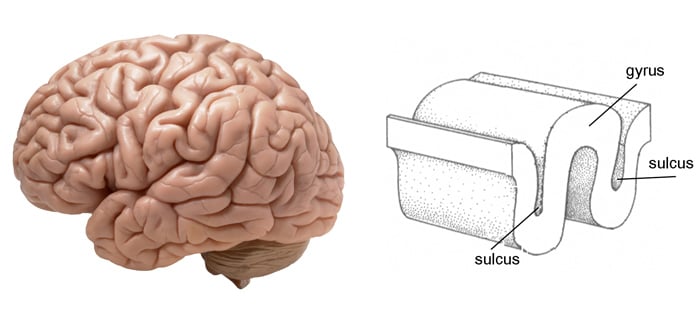
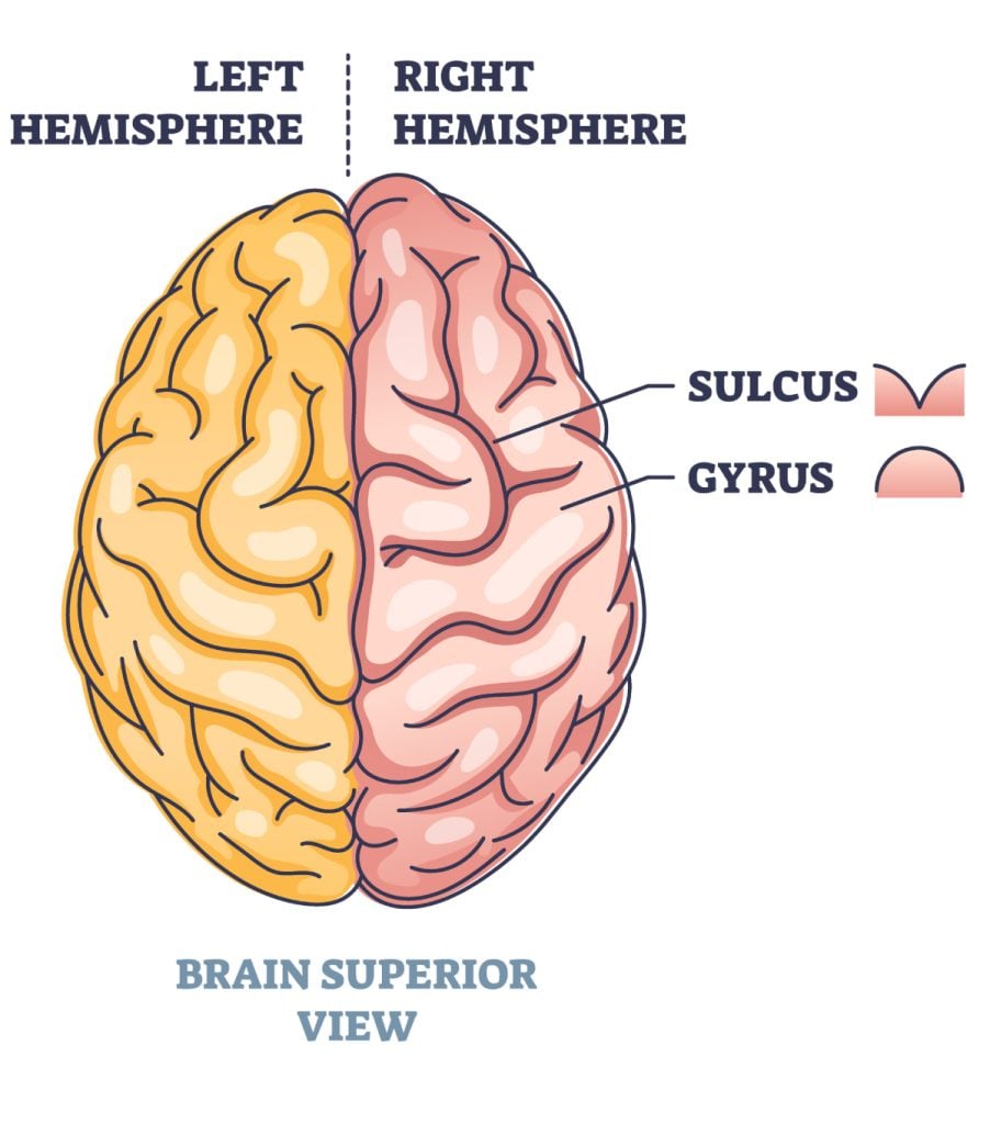
The surface of the brain, known as the cerebral cortex, is very uneven, characterized by a distinctive pattern of folds or bumps, known as gyri (singular: gyrus), and grooves, known as sulci (singular: sulcus).
These gyri and sulci form important landmarks that allow us to separate the brain into functional centers.

Gyri (singular: gyrus) and sulci (singular: sulcus) are the raised and folded structures, respectively, on the cerebral cortex of the brain.
- Gyri (gyrus): These are the raised, convex ridges on the surface of the cerebral cortex. They increase the surface area of the cortex, allowing for greater cognitive processing. Prominent gyri, like the precentral gyrus, are associated with specific functions, such as motor control.
- Sulci (sulcus): These are the grooves or indentations that separate the gyri. Some sulci are deep and define major divisions in the brain, while others are more shallow. Well-known sulci, such as the central sulcus, separate major brain areas like the frontal lobe from the parietal lobe.
Together, the pattern of gyri and sulci gives the brain its characteristic wrinkled appearance, maximizing the cortical surface area within the confines of the skull.
Gyri
The brain has an overall wrinkled appearance, consisting of many ridges and indentations. A gyrus (plural: gyri) is the name given to the bumps and ridges on the cerebral cortex (the outermost layer of the brain).
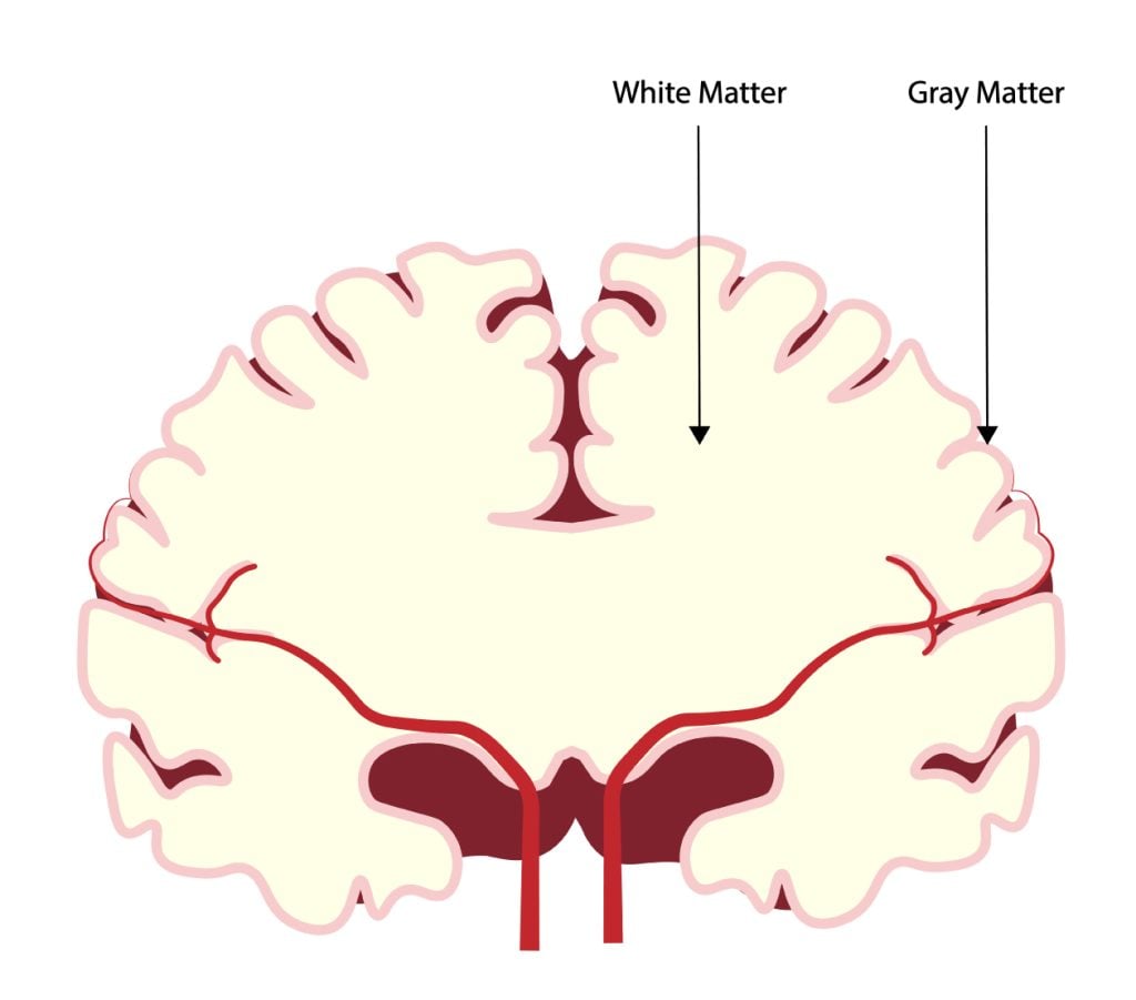
Gyri are found on the surface of the cerebral cortex and are made up of grey matter consisting of nerve cell bodies and dendrites.

They are unique structures that are important as they increase the brain’s surface area. A larger surface area means that more neurons can be packed into the cortex so that it can process more information. Ultimately, cognitive functions will be better with gyri without having to increase the actual brain size, which would not fit into a skull.
The layout and the size of gyri vary from person to person, although there are certain types of gyri that are found in everyone. Although, these types of gyri can vary in size and location between individuals.
There are specific types of gyri that are necessary for the brain’s function. For instance, the precentral gyrus is important as it is the primary motor center of the brain.
Another important area is the superior temporal gyrus which holds Wernicke’s area, an area vital for language development and the comprehension of speech.
As gyri are important to the structure of the brain, they have clinical significance. For example, some abnormalities with gyri can result in disorders such as epilepsy.
Below is a listing of several key gyri in the brain and the divisions they create.
Cingulate gyrus
The cingulate gyrus is a component of the limbic system, consisting of a curved fold that covers the corpus callosum (a bundle of nerve fibers that connects the right and left cerebral hemispheres).
The cingulate gyrus has a role in the processing of emotions and the regulation of behavior. As a result, damage to this area can result in emotional and behavioral disorders. This region is also involved in regulating autonomic motor function.
The cingulate gyrus is often referred to in terms of its divisions: anterior and posterior.
The anterior cingulate gyrus is responsible for emotional processing and the vocalization of emotions, as well as being involved in emotional bonding and attachment, especially between the primary caregiver and child.
This may be due to the fact that this structure has connections to the amygdala, a structure that processes emotions. This gyrus also has connections with areas in the frontal lobes, which are important for speech and motor functions involved with speech production, such as Broca’s Area.
The posterior cingulate gyrus has a role in spatial memory, including the ability to process information relating to the spatial orientation of objects in the environment.
This part of the cingulate gyrus has connections to the parietal and temporal lobes, which allows it to coordinate functions in the areas of movement, orientation, and navigation.
Precentral gyrus function
The precentral gyrus is a part of the brain’s cortex responsible for executing voluntary movements, located in the most posterior position of the frontal lobe, outlining the temporal lobes.
The precentral gyrus is the anatomical location of the primary motor cortex, which is what this gyrus is commonly known as. This gyrus works by creating and organizing a map of the body, known as the homunculus or ‘little man.’
The precentral gyrus is believed to contain the motor control for the torso, arms, hands, fingers, and head. This gyrus also works by controlling the motor movements on the body’s contralateral side, meaning the opposite side to which it is located within the brain.
Superior temporal gyrus
The superior temporal gyrus is an area that contains the auditory cortex, which is responsible for the processing of sounds.
Specific sound frequencies are mapped precisely onto the auditory cortex, forming an auditory map that is similar to the homunculus map of the primary motor cortex.
Some areas of the superior temporal gyrus are responsible for processing combinations of frequencies, whilst others are specialized for processing changes in amplitude or frequency. This gyrus also contains Wernicke’s area, discovered by Karl Wernicke in 1871.
Wernicke’s area is a region important in the comprehension of language as well as having a major role in the ability to produce spoken words.
Sulci
A sulcus (plural: sulci) is another name for a groove in the cerebral cortex. Sulci surround each gyrus, and together, the gyri and sulci help to increase the surface area of the cerebral cortex and form brain divisions.
They form brain divisions by creating boundaries between the lobes, so these are easily identifiable and serve to divide the brain into two hemispheres.

A sulcus is a shallow groove that surrounds a gyrus, whereas sulci that are larger or deeper are given the term fissures.
The longitudinal fissure is the large furrow that divides the two hemispheres into left and right. A smooth-surfaced cortex would only be able to increase to a certain extent. Therefore, sulci in the surface area allow for continued growth, overall increasing brain function.
There are two types of sulci that are formed at different times. The primary sulci (e.g., the central sulcus) are formed independently before birth. Secondary sulci, however, are those formed by other factors other than the growth in adjoining areas of the cortex (e.g., the parieto-occipital sulci).
Sulci can also be defined in terms of their depth. A complete sulcus is a sulcus where the groove is very deep (e.g., the collateral sulcus), whereas an incomplete sulcus is not very deep (e.g., the paracentral sulcus).
Listed below are a number of important brain sulci (fissures) of the cerebrum.
Longitudinal fissure
The longitudinal fissure is a deep furrow located within the center of the brain, separating the left and right hemispheres.
Within this fissure is the corpus callosum, which is a bundle of nerve fibers that connects the left and right hemispheres of the brain to send visual, auditory, and somatosensory information between each half.
Central sulcus
The central sulcus, also known as the sulcus of Rolando, separates the parietal and frontal lobes.
This is an essential sulcus because it defines the boundary between the primary motor cortex and primary somatosensory cortex and between the parietal and frontal lobes.
It is believed that as motor functions develop, the shape of the central sulcus will also change due to the role of this sulcus in separating the motor and sensory cortices.
It has also been suggested that the surface area of the central sulcus can affect the handedness of an individual. A larger central sulcus in the left hemisphere has been found in those who are right-handed, whereas, in left-handed people, this sulcus is larger in the right hemisphere.
Parieto-occipital sulcus
The parieto-occipital sulcus is a deep groove that separates the parietal and occipital lobes of the brain.
This sulcus formed a notch on the external surface of the cortex, which serves as an indicator of where the parietal and occipital lobes lie.
This sulcus is also a secondary sulcus as it forms after birth.
Lateral sulcus
The lateral sulcus is a deep groove that separates the parietal and temporal lobes.
This is also known as the Sylvian sulcus and begins near the forebrain, extending to the lateral surface of the brain, with the insular cortex being located immediately deep within this sulcus.
Abnormal Gyri and Sulci
Within the early stages of development, there can be an abnormal pattern of gyri and sulci which can lead to complications. Abnormal patterns are sometimes caused by disorders of cell migration in the developing cortex.
If gyri do not form properly during development, the cerebral cortex will be smoother than it should be, a condition called lissencephaly. Issues with smooth cerebral cortices can be a factor in the development of epilepsy.
Abnormally large gyri can form, leading to pachygyria, and abnormally small gyri can lead to microgyria. These abnormalities can affect the cerebral cortex as a whole, but they may also be localized to one area, and the conditions can coexist which each other.
For instance, an individual’s brain may have an unusually small gyrus and an unusually large gyrus.
Polymicrogyria is a condition that is characterized by an excessive number of gyri in the brain which develops before birth.
With this condition, the sulci will be abnormally shallow in comparison to typically developed brains. This results in an irregular surface to the cortex and can be localized to a single gyrus or can involve many gyri.
The most common symptom of polymicrogyria is the development of epileptic seizures, with the incidence rate of epilepsy reported to range from between 60-85% of those with polymicrogyria between the ages of 4 and 12 years of age.
Other symptoms of polymicrogyria can include a delay in development, problems with speech and swallowing, muscle weakness, or paralysis. Severe cases of polymicrogyria, named bilateral generalized polymicrogyria, can affect the brain as a whole.
Damage or developmental irregularities with specific gyri or sulci can affect specific functions. Perisylvian syndrome is a rare neurological disease that results from damage to the lateral sulcus (or Sylvian sulcus), resulting in language and speech problems.
Finally, abnormalities of the superior temporal gyrus have been associated with some of the symptoms of schizophrenia (Barta et al., 1990).
It has been suggested that in individuals with one of the positive symptoms of schizophrenia, auditory hallucinations, their superior temporal gyrus has structural and functional deficits.
References
Banker, L., & Tadi, P. (2020). Neuroanatomy, Precentral Gyrus. StatPearls [Internet].
Barta, P. E., Pearlson, G. D., Powers, R. E., Richards, S. S., & Tune, L. E. (1990). Auditory hallucinations and smaller superior temporal gyral volume in schizophrenia. The American Journal of Psychiatry.
Haines, D. E., & Mihailoff, G. A. (2017). Fundamental Neuroscience for Basic and Clinical Applications E-Book . Elsevier Health Sciences.
Spreafico, R., & Tassi, L. (2012). Cortical malformations. Handbook of clinical neurology, 108, 535-557.

