On This Page:
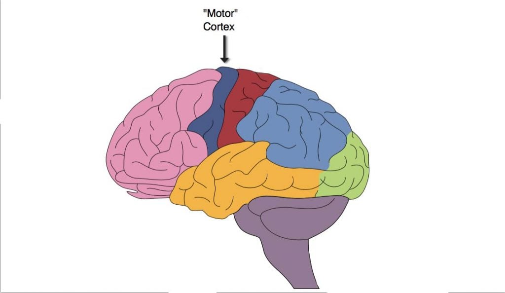
The motor cortex is an area within the brain’s cerebral cortex involved in the planning, control, and execution of voluntary movements.
It is located in the frontal lobe and works with other brain areas and the spinal cord to translate thought into physical motion. In psychology, the motor cortex is studied for its role in skills acquisition, muscle coordination, and the integration of sensory information to produce complex motor actions.
The motor cortex can be divided into the primary motor cortex and the nonprimary motor cortex.
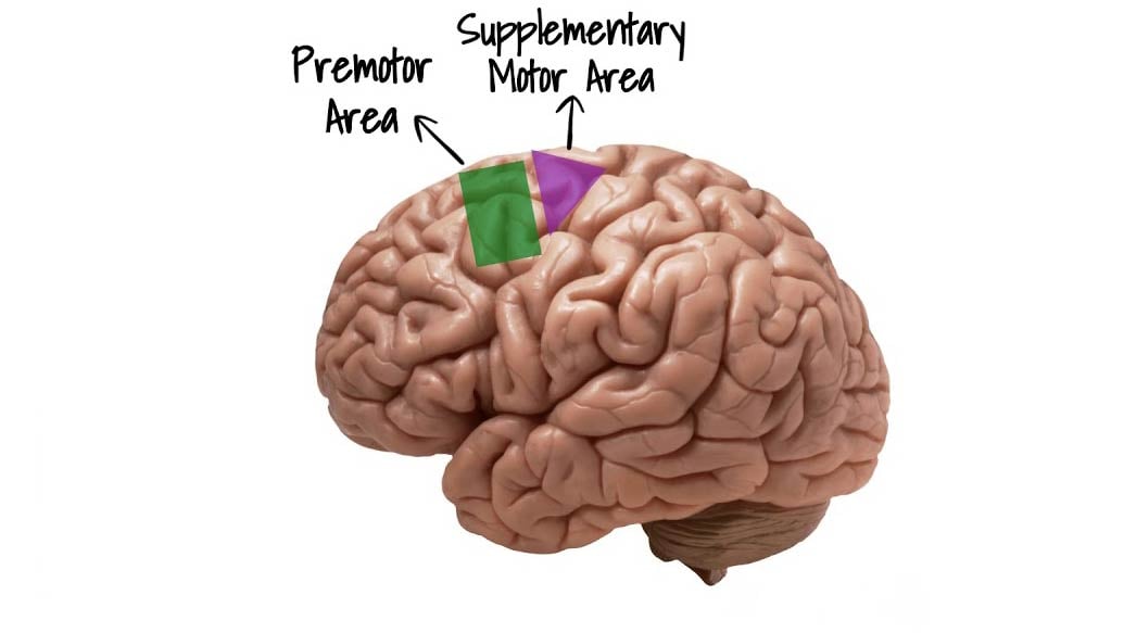
The primary motor cortex is critical for initiating motor movements. The nonprimary motor cortex, further divided into other areas such as the premotor and supplementary cortex, involves planning, initiating, and selecting the correct movement.
Both hemispheres have a motor cortex, with each side controlling muscles on the opposite side of the body (i.e., the left hemisphere controls muscles on the right side).

Different areas of the motor cortex control different parts of the body, and these are in the same sequence as in the body (e.g., the part of the cortex controlling the foot is next to the part controlling the leg, etc.)
The central sulcus is a groove that runs down the side of the cerebral hemispheres between the frontal and parietal lobes. Specifically, the primary motor cortex is found in a gyrus (ridge) called the precentral gyrus, which is positioned just in front of the central sulcus.
The nonprimary motor cortex, however, is located anterior to the primary motor cortex. The motor cortex is the only motor control center above the spinal cord, which can directly communicate with most of the other motor control structures such as the thalamus, basal ganglia, brain stem, and spinal cord.
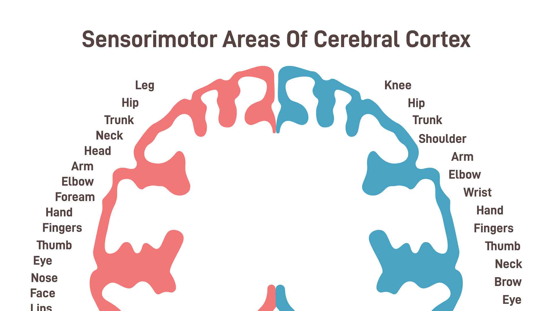
This region also contains an inverted representation of the opposite half of the body, known as a motor homunculus. Therefore, the top part of the cortex stimulates movements of the leg, whereas the movements of the face are stimulated by the lowest part of the motor cortex.
Thus, each movement of the body is represented by neuronal activity in different areas of the cortex. This is similar to the sensory homunculus of the sensory cortex, which is situated immediately behind the motor cortex.
Motor Cortex Functions
Primary Motor Cortex
The primary motor cortex is a region of the motor cortex that is important for initiating motor movements.
The areas of the primary motor cortex correspond precisely to specific body parts. The primary motor cortex contains large neurons (nerve cells) with triangular-shaped bodies called pyramidal neurons.
Pyramidal neurons, also known as upper motor neurons, and the primary output cells of the motor cortex. The axons of these neurons exit the motor cortex, carrying information about voluntary movements it wishes to make.
To do this, the pyramidal neurons enter one of the tracts of the pyramidal system: either the corticospinal or corticobulbar tracts. When pyramidal neurons travel through the corticospinal tract, it carries with it information to the spinal cord.
Once the information has reached the spinal cord, it can then be used to initiate movements. In contrast, the pyramidal neurons, which travel down the corticobulbar tract, carry motor information to the brain stem at the base of the brain.
Once here, cranial nerve nuclei are stimulated to initiate movements of the head, neck, and face. Both of these tracts, therefore, carry specific information about voluntary movements down from the brain.
The primary motor cortex does not generally control muscles directly but tends to initiate individual movements or sequences of movements that rely on the activity of many muscle groups.
To directly supply skeletal muscles to cause movement, the pyramidal (or upper) neurons form connections with other neurons called lower motor neurons, which are efferent neurons that connect the central nervous system to the muscles.
Nonprimary Motor Cortex
The nonprimary motor cortex is further divided into two areas: the premotor cortex and the supplementary motor cortex. The premotor cortex is thought to be involved in planning and executing motor movements.  It is also thought to contain mirror neurons, which are a class of neurons that regulate activity both when individuals perform specific motor acts as well as when they observe the same or similar action performed by another individual.
Because mirror neurons are present in this region, the premotor cortex is also thought to play a role in learning, particularly in the imitation of others.
The premotor cortex is thought to use information from other cortical regions, such as the cerebellum, in order to select appropriate movements.
The supplementary motor cortex is thought to be critical to the execution of sequences of movement, the execution of motor skills, and the control of movement. This can involve taking a role in deciding to change to a different movement based on sensory input.
Motor Homunculus
The motor homunculus is a representation of the body parts along the primary motor cortex, or precentral gyrus.
Each part of the body that can move is represented along this gyrus in an anatomical fashion, representing the contralateral side of the body.
This means that the primary cortex in the right cerebral hemisphere represents motor movements on the left side of the body and vice-versa.  The body parts are organized in a way that the lowest parts of the body are arranged near the top of the precentral gyrus, whilst the top of the body is arranged near the lower parts of the gyrus.
Thus, stimulation of the anterior precentral gyrus would elicit movements of the contralateral leg. Going along the gyrus, the trunk and arm movements would be a result of activity in this area, followed by movements of the hand and fingers.
Finally, near the bottom of the gyrus, movements of the face, eyes, tongue, and jaw would result from activity in this area.
The representations of the body parts which perform more movements, or more precise movements, such as the hands and face, are disproportionately large compared to representations of other body parts that can only perform a few or less refined movements, such as the trunk and legs.
Therefore, the reason for the distorted representations of the homunculus is not due to how large in size the parts of the body are but is due to how richly supplied those parts are by the motor cortex.
The motor homunculus is not to be confused with sensory homunculus, which is a sensory representation in the somatosensory cortex in the postcentral gyrus.
This homunculus represents how body parts feel, whereas the motor homunculus represents how body parts move.
The regions of the body representations on the sensory homunculus differ slightly from that of the motor counterpart as some areas of the body are more sensitive to sensations rather than movements, such as the head.
Motor Cortex Dysfunction
Common causes of dysfunction of the motor cortex are as a result of a stroke or traumatic brain injury. Damage to this area may result in dysfunctions associated with the pyramidal neurons, a condition known as upper motor neuron disease.
Below are some of the symptoms that are associated with upper motor neuron disease:
- Weakness of one side of the body – if damage has occurred on the motor cortex in the left hemisphere, this can result in weakness on the right side of the body. This may mean that individuals may struggle to move or raise their arm, or one side of the face may appear drooped.
- Decreased motor control – damage to the motor cortex could result in poorer coordination of motor movements and poorer dexterity.
- Depending on the damage, this could negatively affect the fine motor skills of an individual. Fine motor skills are precise motor movements such as writing or fastening buttons on a shirt.
- Overactive reflexes – sometimes, individuals with upper motor neuron disease will perform involuntary reactions of their muscles, including muscle stretch reflexes. These can often be exaggerated and can make it difficult to perform precise movements.
- Decreased endurance – because of damage, the muscles may become fatigued more easily and quicker than normal.
- Altered muscle tone – the muscle tone can be either abnormally high or low. This can have a direct affect on mobility as well as coordination of movements.
Although it is not possible to repair damaged neurons of the motor cortex, muscle control functions can be recovered after damage to this area of the brain.
The brain has the ability for neuroplasticity, which is the ability of the brain to reorganize neurons and compensate for damaged areas.
Thus, the brain can change its structure slightly so that parts of the brain that are healthier can take control of muscle movements in place of the motor cortex.
Neuroplasticity is often an aim in physical and occupational therapies for individuals with motor cortex damage. This ability can usually be triggered through repetitive exercise and activities with the therapist to activate a particular muscle group.
The more this is practiced, the more those muscle pathways will be reinforced. Eventually, new pathways will be established so individuals can perform movements easier.
References
Purves, D., Augustine, G., Fitzpatrick, D., Katz, L., LaMantia, A., McNamara, J., & Williams, S. (2001). Neuroscience 2nd edition. Sunderland (ma) sinauer associates. Types of Eye Movements and Their Functions.
Neuroscientifically Challenged (2015, October 23). Know Your Brain: Motor Cortex. https://www.neuroscientificallychallenged.com/blog/know-your-brain-motor-cortex
Knierim, J. (2020, October 20). Chapter 3: Motor Cortex. Neuroscience Online. https://nba.uth.tmc.edu/neuroscience/m/s3/chapter03.html
Flint Rehab (2020, November 19). Primary Motor Cortex Damage: Definition, Symptoms, and Treatment. https://www.flintrehab.com/primary-motor-cortex-damage/

