On This Page:
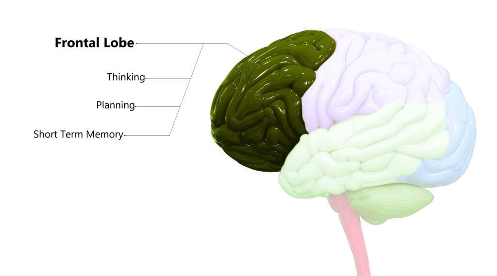
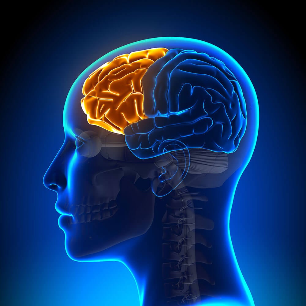
The frontal lobe is located behind the forehead, at the front of the brain. These lobes are part of the cerebral cortex and are the largest brain structure.
The frontal lobe’s main functions are typically associated with ‘higher’ cognitive functions, including decision-making, problem-solving, thought, and attention.
It contains the motor cortex, which is involved in planning and coordinating movement; the prefrontal cortex, which is responsible for higher-level cognitive functioning; and Broca’s Area, which is essential for language production

Frontal Lobe Functions
Below is a list of some of the associated functions of the frontal lobe:
- Executive processes (capacity to plan, organize, initiate, and self-monitor)
- Voluntary behavior
- Problem-solving
- Voluntary motor control
- Intelligence
- Language processing
- Language comprehension
- Self-control
- Emotional control
The frontal lobes are believed to be our behavior and emotional control centers, meaning that this area is activated when needing to control our behaviors to be socially appropriate and to control our emotional responses, especially in social situations.
Moreover, the frontal lobes are thought to be the home of our personalities.
Alike to most lobes in the brain, there are two frontal lobes located in the left and right hemispheres.
Each lobe controls the operations on opposite sides of the body: the left hemisphere controls the right side of the body and vice versa.
It is believed the left frontal lobe is the most dominant lobe and works predominantly with language, logical thinking, and analytical reasoning.
The right frontal lobe, on the other hand, is most associated with non-verbal abilities, creativity, imagination, and musical and art skills.
The frontal lobe, like other structures of the brain, does not always work in isolation from each other. The frontal lobes work alongside other brain regions in order to control a variety of functions.
Substructures
The frontal lobe contains the motor cortex, which is involved in planning and coordinating movement; the prefrontal cortex, which is responsible for higher-level cognitive functioning; and Broca’s area, which is essential for language production.
Prefrontal Cortex
The prefrontal cortex is primarily responsible for the ‘higher’ brain functions of the frontal lobes, including decision-making, problem-solving, intelligence, and emotion regulation.
This area has also been found to be associated with the social skills and personality of humans.
This idea is supported by the famous case study of Phineas Gage, whose personality changed after losing a part of his prefrontal cortex after an iron rod impaled his head.
The frontal cortex has also been shown to be activated when an experience becomes conscious. Different ideas and perceptions are bound together in this region, both of which are necessary for conscious experience. Concluding that this area may be especially important for consciousness.
Cognitive disorders that have been shown to be linked to this region are attention deficit hyperactivity disorder (ADHD), Autism, bipolar disorder, depression, and schizophrenia.
The prefrontal cortex can be further divided into the dorsolateral prefrontal cortex and the orbitofrontal cortex.
Motor and Premotor Cortex
The motor cortex is critical for initiating motor movements, as well as coordinating motor movements, hence why it is called the motor cortex.
Each area of the motor cortex corresponds precisely with specific body parts. For instance, there is an area that controls the left and the right foot.
The premotor cortex is associated with planning and executing motor movements. Within this area, voluntary movement is rehearsed, distinguishing these movements from unconscious reactions.
The premotor cortex has also been shown to be important for imitation learning through the use of mirror neurons. These neurons essentially allow us to reflect the body language, facial expressions, and emotions of others.
Furthermore, the prefrontal cortex can support cognitive functions of a social kind, such as showing empathy.
Broca’s Area
Another region of the frontal lobes worth mentioning is Broca’s area. This region is located in the dominant hemisphere of the frontal lobes, which is the left side for around 97% of humans.
This region is associated with the production of speech and written language, as well as with the processing and comprehension of language.
The name is taken from the French scientist Paul Broca, whose work with language-impaired patients led him to conclude that we speak with our left brains.
Language differences in those with Autism may be correlated to differences in the structure and function of Broca’s area (Bauman & Kemper, 2005).
Damage
As the frontal lobes are situated at the front of the brain and are large in size, this makes them more susceptible to damage. This area is the most common for traumatic brain injuries, with damage to this region causing a variety of symptoms.
Below is a list of symptoms that may occur if an individual has experienced damage within their frontal lobe:
- Paralysis
- Changes in mood
- Attention deficits
- Atypical social skills
- Difficulty problem-solving
- Lack of impulse control/ risk-taking
- Loss of spontaneity in social interactions
- Reduced motivation
- Impaired judgment
- Reduced creativity
Damage to Broca’s area, in particular, has been shown to affect the ability to speak, understand language, and produce coherent sentences.
One of the most famous case studies associated with frontal lobe damage is the case of Phineas Gage. He was a railway construction worker who suffered an unfortunate accident when a metal rod impaled his brain in the frontal region.
Gage survived this accident but was said to have experienced some personality changes because of the trauma. Before the accident, Gage was described as a ‘well-balanced’ and smart, energetic person.
After his accident, he was described as being childlike in his intellectual capacities and had a loss of social inhibition (behaving in ways that were considered socially inappropriate).
This case study implies that the frontal lobes are essential to our personalities, intelligence, and social skills. As well as trauma to the head is a cause of damage to the frontal lobes, there are many other causes that can lead to damage.
For instance, a brain tumor, stroke, or infection can cause deficits in this lobe. Similarly, conditions such as cerebral palsy, Huntington’s disease, dementia, or other neurodegenerative diseases can lead to associated damage.
If someone is suspected of having frontal lobe damage, there are methods to diagnose this. Magnetic resonance imaging (MRI) and computerized tomography (CT) scans can detect some differences in the frontal lobes after suffering a stroke or infection, as well as be able to detect dementia.
Also, neuropsychological evaluations can be completed to test for areas such as speech comprehension, social behavior, memory, problem-solving, and impulse control, among others.
One common test to establish frontal lobe damage is the Wisconsin Card Sorting task. Within this task, individuals will be shown cards of varying sorts, such as some having symbols, numbers, different shapes, and colors on them.
They will then be asked to sort the cards by a certain criterion, which will then change throughout the test. Those who have damage to a certain part of the frontal lobes may struggle with this task and will not adjust to new sorting criteria. They will stick with the original criteria (this is called perseveration).
Other tests worth noting as finger tapping tests, to test for motor skill ability, and the Token Test, which tests for language skills.
To be able to treat frontal lobe damage, occupational, speech, and physical therapy can be helpful for rehabilitating these lost or damaged skills.
Finally, a talking therapy called cognitive behavioral therapy (CBT) is common for working on regulating emotions and aiding impulse behaviors.
CBT may not fully treat physical damage to the frontal lobes, but it can help those with impairments cope and manage their symptoms.
Research Studies
- Semmes, Weinstein, Ghent & Teuber (1963) suggested that the frontal lobes played a part in spatial orientation, particularly our body’s orientation in space.
- Eslinger & Grattan (1993) investigated damage to the prefrontal cortex. They suggested that people with damage to this area may not have problems with word comprehension or identifying objects by their names, but if asked to say or write as many words as possible or describe as many uses of an object, they would find this task difficult. This shows that damage to one area associated with language does not impair all aspects of language.
- Kolb & Milner (1981) discussed the involvement of the frontal lobes in facial expressions. They found that patients with frontal lobe damage had difficulty expressing spontaneous facial expressions and would also show fewer facial movements spontaneously.
- Kaufman, Geyer & Milstein (2017) reported that patients who suffered damage to their frontal lobes had changes to their personalities.
- It was found that these patients developed an abrupt, suspicious, and sometimes even argumentative manner.
- Some patients were reported to have displayed ‘emotional incontinence,’ whereby they would have bouts of pathological laughing or crying.
- Walker & Blummer (1975) found that damage to the frontal lobes resulted in displays of abnormal sexual behavior in the orbital region and reduced sexual interest if the dorsolateral region was damaged.
- Stuss et al. (1992) found that damage to certain areas of the frontal lobes resulted in ‘bland’ personalities. These patients also displayed fewer signs of distress in emotionally heightened situations.
- Catani et al. (2016) investigated the brains of people with Autism and found support for the hypothesis that Autism is associated with different connectivity in the frontal lobe region compared with neurotypical individuals.
- Mubarik & Tohid (2016) conducted a literature review of studies that investigated the frontal lobes of those with schizophrenia.
- They found that many people with schizophrenia have differences in the structure of white matter, grey matter, and functional activity in their frontal lobes compared to those without the condition.
References
Bauman, M. L., & Kemper, T. L. (2005). Neuroanatomic observations of the brain in autism: a review and future directions. International Journal of Developmental Neuroscience, 23(2-3), 183-187.
Catani, M., Dell’Acqua, F., Budisavljevic, S., Howells, H., Thiebaut de Schotten, M., Froudist-Walsh, S., … & Murphy, D. G. (2016). Frontal networks in adults with autism spectrum disorder. Brain, 139(2), 616-630.
Eslinger, P. J., & Grattan, L. M. (1993). Frontal lobe and frontal-striatal substrates for different forms of human cognitive flexibility. Neuropsychologia, 31(1), 17-28.
Kaufman, D. M., Geyer, H. L., & Milstein, M. J. (2017). Chapter 21-neurotransmitters and drug abuse. Kaufman’s Clinical Neurology for Psychiatrists. 8th ed. Amsterdam: Elsevier, 495-517.
Kolb, B., & Milner, B. (1981). Performance of complex arm and facial movements after focal brain lesions. Neuropsychologia, 19(4), 491-503.
Mubarik, A., & Tohid, H. (2016). Frontal lobe alterations in schizophrenia: a review. Trends in Psychiatry and Psychotherapy, 38(4), 198-206.
Semmes, J., Weinstein, S., GHENT, G., Meyer, J. S., & Teuber, H. L. (1963). Correlates of impaired orientation in personal and extrapersonal space. Brain, 86 (4), 747-772.
Stuss, D. T., Ely, P., Hugenholtz, H., Richard, M. T., LaRochelle, S., Poirier, C. A., & Bell, I. (1985). Subtle neuropsychological deficits in patients with good recovery after closed head injury. Neurosurgery, 17 (1), 41-47.
Walker, A. E., & Blumer, D. (1975). The localization of sex in the brain. In Cerebral localization (pp. 184-199). Springer, Berlin, Heidelberg.


