On This Page:
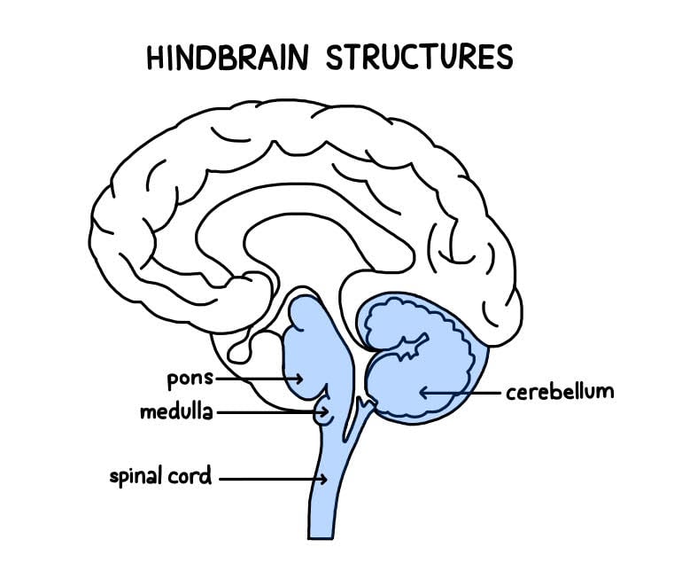
The hindbrain is located at the lower back part of the brain and includes most of the brainstem (containing the medulla and pons), and the cerebellum. The hindbrain is located at the back of the head and looks like an extension of the spinal cord.

The hindbrain is also known as the rhombencephalon and is one of the most crucial parts of the central nervous system (CNS) as it connects the brain to the spinal cord so that messages can be sent from the brain, down the spinal cord, to the rest of the body.
The hindbrain is essentially an extension of the spinal cord, with tracts of axons running through the spinal cord to the hindbrain, which integrates the incoming sensory information and coordinates motor responses.
Hindbrain function
The hindbrain’s chief role is to coordinate the vital functions of our bodies, such as breathing and heart rate. Therefore, the hindbrain is important for survival.
Another main function of the hindbrain is the organization of motor reflexes, mostly controlled by the cerebellum structure. Similarly, the hindbrain is responsible for sleep activity and wakefulness.
Below are the hindbrain parts and their functions.
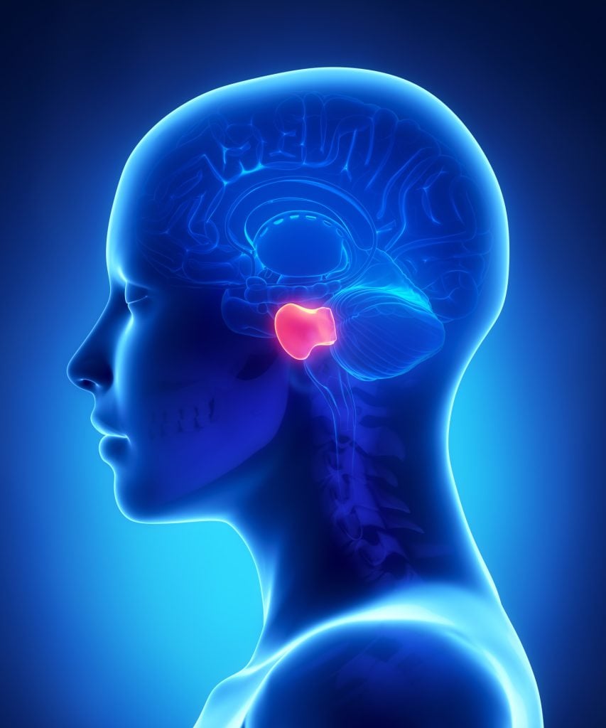
Pons
The pons comes from the Latin word for bridge, named so, as it essentially forms a bridge from the brainstem to the cerebral cortex.
The pons is situated right underneath the midbrain and serves as the coordination center for signals which flow between the two cerebral hemispheres and the spinal cord.  This structure is strongly associated with many autonomic functions, such as breathing, taste, sleeping, and circuits that generate respiratory rhythms.
The pons is also involved in analyzing sensory data and is where auditory information enters the brain.
Within the pons are four types of cranial nerves – these are nerves that help control head muscles and receive sensory information from the head:
- Abducens nerve – these nerves coordinate eye movements.
- Facial nerves are responsible for coordinating the movement and sensations in the face.
- Vestibulocochlear nerve – these process sounds coming into the brain and aid in maintaining balance.
- Trigeminal nerve – this help to coordinate the action of chewing and carrying sensory information from the face and the head.
The pons also contain cell groups that are responsible for transferring signals from the cerebrum to the cerebellum.
Some of these cell groups are part of the reticular formation, which is a network of neurons extending throughout the brainstem with the job of regulating alertness, sleep, and wakefulness.
The nuclei of the pons (pontine nuclei) are involved in learning and remembering motor skills. These nuclei act as relay stations for nerve signals from the motor cortex, which travel to the cerebellum behind the pons.
Cerebellum
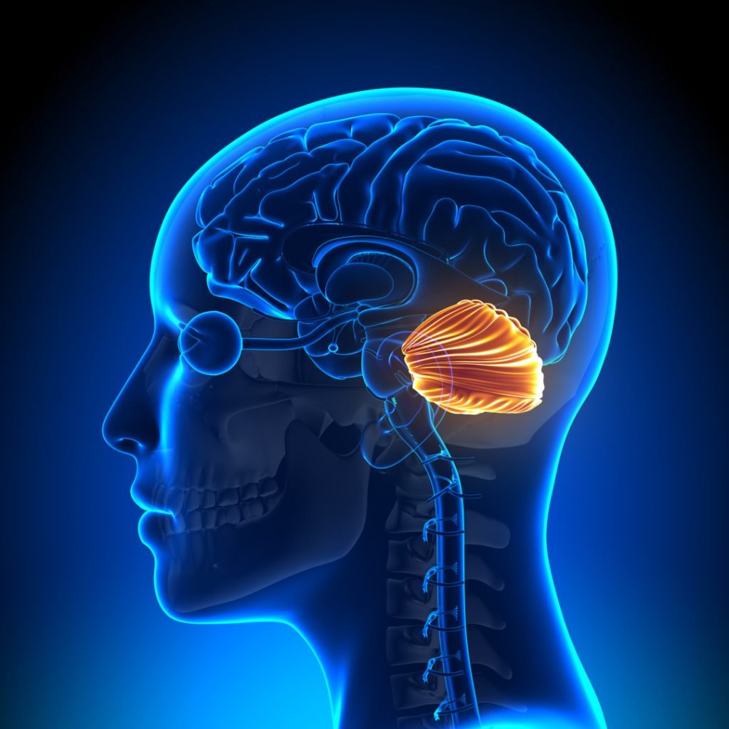
The cerebellum, Latin for ‘little brain’, is a structure of the hindbrain located behind the pons and brainstem.  The cerebellum’s main role is to monitor and regulate motor behavior, particularly automatic movements and balance.
It was once believed that the cerebellum was only involved in coordinating motor movements. We now, however, understand that the cerebellum plays a much bigger role in a variety of functions and communicates signals to other areas of the brain.
The cerebellum has also been found to be involved in motor learning, sequence learning, reflex memory, mental function, and emotional processing.
Although the cerebellum only accounts for 10% of the overall brain mass, it contains over half of the nerve cells than the rest of the brain combined.
In particular, the cerebellum contains cells called Purkinje cells. These are some of the largest neurons within the human brain and have highly complex dendrite branches (nerve endings), which means it is capable of processing many signals at once.
Medulla
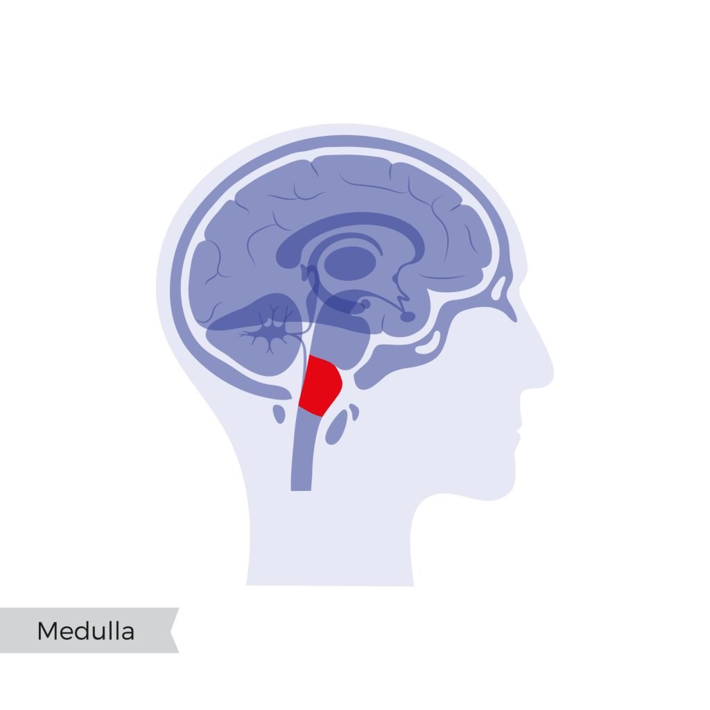
The medulla is a long stem-like structure of the hindbrain, which makes up the lowest part of the brainstem, lying next to the spinal cord.  The medulla controls many functions outside of conscious control, such as breathing, blood flow, blood pressure, and heart rate. This makes the medulla a vital structure for survival.
This structure also involves many involuntary reflexes, such as coughing, sneezing, and swallowing. The medulla contains four types of cranial nerves within it:
- Glossopharyngeal nerve – these nerves coordinate some taste sensations as well as movements of the mouth.
- Vagus nerve – these also control mouth movements, as well as our voice and gag reflexes.
- Accessory nerve – these coordinate movements of the head and the neck.
- Hypoglossal nerve – these nerves control movements of the tongue and muscles involved in speech.
The medulla transmits signals between the spinal cord and higher brain levels and housing nuclei that are centers for automatic and involuntary behaviors.
Damage to the Hindbrain
Symptoms or conditions associated with damage to the hindbrain depend on the structure which is damaged.
Damage to the pons is associated with symptoms such as impaired breathing, sleep disturbances, loss of taste, loss of muscle function (except eye movement), and deafness.
In worse cases, this could result in paralysis or death. A type of stroke called pontine stroke is limited to the pons and can lead to paralysis.
A rare condition associated with damage to an area of the pons is called ‘Locked-in Syndrome’. If an individual has this condition, they are aware of their surroundings and can see and hear.
However, they cannot activate any voluntary muscles that are under conscious control, so they will be unable to move or react. This condition can result from an injury or a lack of blood supply whilst experiencing a stroke.
If the brainstem gets damaged, this can result in difficulties with balance and moving, dizziness, and lack of motor function. The brain stem is also associated with sleep disorders such as insomnia and sleep apnea.
In particular, if the medulla becomes damaged, this can lead to respiratory failure, paralysis, or loss of sensation. Damage to this structure can be fatal as it helps the heartbeat. Therefore, damage to this area can cause termination in a heartbeat, resulting in death.
Finally, cerebellar damage results in the breakdown and destruction of nerve cells which can have long-last effects.
Symptoms associated with damage are a loss of fine coordination, being unbalanced, experiencing tremors, inability to walk, vertigo, and slurred speech. Drinking alcohol has an immediate and temporary effect on the cerebellum as the body’s coordination and movements become clumsy.
Someone who has drunk a lot of alcohol may not be able to walk in a straight line and may have trouble balancing. Repeated alcohol misuse can become an alcohol use disorder and can have long-lasting impacts on the cerebellum and lead to these symptoms being more long-lasting.
Other causes of damage to the cerebellum can come from injury to the head, such as falling backward and hitting the back of the head where the cerebellum lies. Brain tumors and infections in the brain can also cause long-lasting damage to the cerebellum.
Damage could also occur through medical issues such as having Parkinson’s disease, multiple sclerosis, and experiencing a stroke.
Although some diseases and conditions associated with damage to the hindbrain cannot be avoided or the cause is unknown, there are lifestyle choices that can be taken to preserve the health of the brainstem, specifically the cerebellum.
Limiting or stopping smoking, as well as limiting the amount of alcohol consumption, is a suggestion. This is because they both contribute to raising blood pressure which could ultimately lead to a stroke.
Drinking alcohol can also result in being unbalanced, which can be a higher risk of injury. Exercising more and eating a healthy diet both help lower blood pressure and, thus, the risk of a stroke, so this is encouraged to protect the brain.
Finally, protecting the head in general, such as wearing helmets when cycling, wearing seatbelts in the car, and taking care to prevent falls at home, can limit the risk of damage to the structures of the hindbrain.
Research Studies
- Several studies have investigated the hindbrain, its functions, and its links with cognitive disorders. Kolb (1987) suggested that some of the symptoms associated with posttraumatic stress disorder (PTSD) are the result of neuronal impairments within the hindbrain structures, especially those areas concerned with aggressive expression and the sleep and dream cycle.
- A magnetic resonance imaging (MRI) study which investigated the brains of those who were diagnosed as Autistic and those who were neurotypically found decreased grey matter volumes in the brainstems of Autistic individuals (Jou et al., 2009).
- As grey matter is associated with processing information due to the number of neurons there, this suggests that perhaps Autistic individuals do not process as much information or process information differently from those who are not Autistic.
- A group of researchers investigated the heart and respiratory function in those who had been diagnosed with Parkinson’s disease.
- They found that the medulla being damaged in these patients may have been the underlying reason for the heart and breathing problems with these patients (Pyatigorskaya et al., 2016).
- During normal resting eye movement (REM) sleep, the body becomes paralyzed, preventing us from acting out movements during sleep. Xi and Luning (2008) investigated people who had REM sleep behavior disorder as a result of a deficit in the brainstem.
- This is characterized by a loss of this paralysis, which can result in disruptive behaviors during sleep. This can include screaming, thrashing about, kicking, and punching.
- Within the hindbrain, the cerebellum is the most researched structure. Stoodley (2016) suggested that dysfunction of the cerebellum is found in individuals with developmental disorders such as Autism, attention deficit hyperactivity disorder (ADHD), and developmental dyslexia.
- There also seems to be a reduction in the number of Purkinje cells within the cerebellum of those with Autism (Whitney et al., 2008). As the Purkinje cells can send many signals around the brain at once, this suggests that those with Autism have a deficit in sending signals compared to neurotypical brains.
- Another study of the cerebellum has shown that injuries or strokes to this area have shown many deficits in language processing, visuospatial abilities, executive function, and attention (Tedesco et al., 2011).
References
Jou, R. J., Minshew, N. J., Melhem, N. M., Keshavan, M. S., & Hardan, A. Y. (2009). Brainstem volumetric alterations in children with autism. Psychological Medicine, 39 (8), 1347.
Kolb, L. C. (1987). A neuropsychological hypothesis explaining posttraumatic stress disorders. The American Journal of Psychiatry, 144, 989-95.
Pyatigorskaya, N., Mongin, M., Valabregue, R., Yahia-Cherif, L., Ewenczyk, C., Poupon, C., Debellemaniere, E., Vidailhet, M., Arnulf, I. & Lehéricy, S. (2016). Medulla oblongata damage and cardiac autonomic dysfunction in Parkinson disease. Neurology, 87 (24), 2540-2545.
Stoodley, C. J. (2016). The cerebellum and neurodevelopmental disorders. The Cerebellum, 15 (1), 34-37.
Tedesco, A. M., Chiricozzi, F. R., Clausi, S., Lupo, M., Molinari, M., & Leggio, M. G. (2011). The cerebellar cognitive profile. Brain, 134 (12), 3672-3686.
The University of Queensland. (n.d). The hindbrain. Retrieved April 29, 2021, from https://qbi.uq.edu.au/brain/brain-anatomy/hindbrain
Whitney, E. R., Kemper, T. L., Bauman, M. L., Rosene, D. L., & Blatt, G. J. (2008). Cerebellar Purkinje cells are reduced in a subpopulation of autistic brains: a stereological experiment using calbindin-D28k. The Cerebellum, 7 (3), 406-416.
Xi, Z., & Luning, W. (2009). REM sleep behavior disorder in a patient with pontine stroke. Sleep medicine, 10 (1), 143-146.

